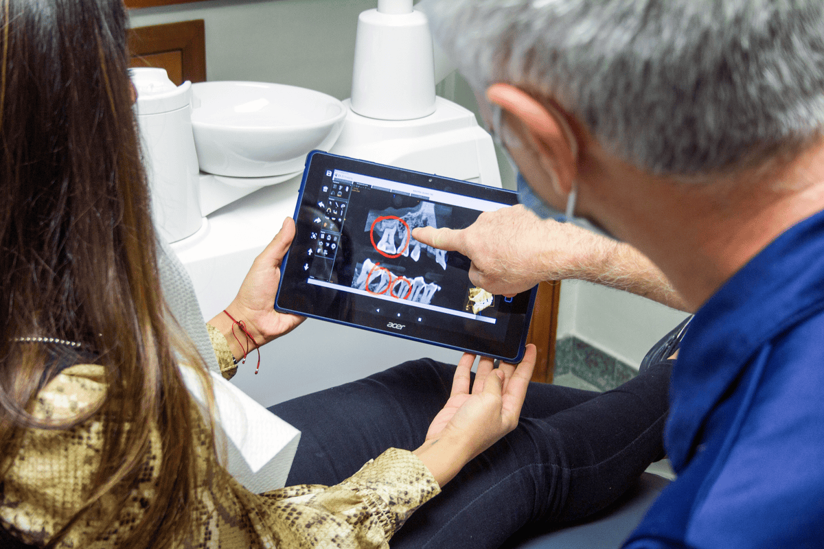
Diag image is a cornerstone of modern healthcare. It allows clinicians to view the inside of the human body with unprecedented clarity and precision. Medical imaging workstations continue to play a central role in the clinical workflow, enabling clinicians to make accurate diagnoses and improve patient care. This is the power of diagnostic imaging technology.
According to healthcare industry studies, approximately 12 million Americans are affected by diagnostic errors each year, with imaging-related errors accounting for a significant proportion. To address this need, advanced diagnostic imaging technology offers radiologists and clinicians effective solutions for improving diagnostic accuracy and reducing interpretation errors.
What is diagnostic imaging?
Diag Image technology, often simply referred to as diagnostic imaging, encompasses the techniques and equipment used to create visual representations of the internal structures of the human body. Doctors can use these images to diagnose, monitor, and treat various conditions without invasive procedures.
Studies show that approximately 12 million people in the United States are affected by diagnostic errors each year, with imaging-related errors accounting for a significant proportion of these. Essentially, using imaging techniques is like looking through a window into the body. It allows for a clear view of soft tissues, bones, and the blood vessels behind them.
Common Imaging Techniques
Overview of the most commonly used Diag Image techniques:
X-rays
These electromagnetic rays produce images of bones and other tissues in the body. X-rays help detect fractures, infections, and tumors.
Computed Tomography (CT)
This technique combines multiple X-ray images taken from different angles to create cross-sectional images of the body. CT scans provide detailed images of bones, blood vessels, and soft tissues. This allows for the diagnosis of cancer, cardiovascular disease, and traumatic injuries.
Magnetic Resonance Imaging (MRI)
MRI scanners use radio frequency signals and strong magnetic fields to create detailed images of organs and tissues. MRI is suitable for imaging soft tissues, joints, the brain, and the spinal cord.
Ultrasound
Ultrasound uses high-frequency sound waves to create images of the inside of the body. In obstetrics, it is used to monitor fetal development and to examine the growth of organs such as the heart, liver, and kidneys.
Positron Emission Tomography (PET)
In PET, a small amount of radioactive material is injected into the body to examine areas of high chemical activity. PET scans are used in oncology for cancer detection and to monitor the progress of therapy.
How Diagnostic Imaging Enables Early and Accurate Diagnosis
Suppose a patient is suffering from persistent headaches. In addition to the symptoms, the doctor may order an MRI. The resulting image can reveal a brain lesion that may indicate a problem such as a tumor or aneurysm at an early stage (before it becomes life-threatening).
Accuracy and Speed
Time is critical in medicine (and healthcare). Take stroke treatment, for example: Every minute without treatment leads to the death of brain cells. Imaging techniques can detect brain hemorrhages or blood clots within minutes, enabling the fastest possible treatment.
Minimally Invasive Yet Informative
Before the development of modern imaging techniques, surgical exploration was the only way to determine the cause of disease. Today, medical imaging offers the same clarity without a single incision. This means fewer risks, faster recovery, and greater patient safety.
Advanced Visualization and Measurement Tools
Diag Image supports specialized measurement tools and features tailored to various imaging modalities. Goniometry measurements, pan and zoom, video inversion, and high-quality image processing contribute to increased diagnostic accuracy across a wide range of clinical applications.
Tool Category: Key Features: Clinical Applications:
Measurement Tools: Goniometry, distance and area calculations; orthopedic scans, cardiac measurements
Image Processing: Pan, zoom, video inversion, contrast adjustment; general radiology, emergency imaging
Visualization Protocols: Automated visualization protocols, layout optimization
Pathology Indexing: Image cataloging, teaching database; teaching, research, case documentation
These tools are particularly valuable for complex cases requiring detailed measurements or comparative analysis. Radiologists can perform precise calculations and annotations directly on the diagnostic interface, streamlining the reporting process.
The Technology Behind Diagnostic Imaging Systems: While it may seem simple—a machine takes an image and displays it on a screen—the science behind diagnostic imaging technology is highly advanced.
Digital Imaging and Artificial Intelligence (AI)
Modern diagnostic imaging systems operate digitally. This means that results can be stored, optimized, and shared across various healthcare platforms. Artificial intelligence (AI) is being gradually integrated into diagnostic systems, enabling radiologists to detect abnormalities such as lung nodules or breast cancer with greater accuracy and efficiency than ever before.
A 2020 study published in The Lancet Digital Health revealed that AI systems match or even surpass radiologists in interpreting diagnostic images for breast cancer screening.
3D Imaging and Reconstruction
New advancements enable the acquisition of 3D images. This allows physicians to rotate and zoom in on images and even simulate surgical planning based on the patient’s actual anatomy. This is especially useful for orthopedic, cardiovascular, and oncological procedures.
The Role of Imaging Centers in Healthcare
In the United States, imaging centers have become hubs of innovation and points of contact for improving access to healthcare. These centers specialize in performing various diagnostic imaging procedures and often offer more specialized care and shorter wait times than hospitals.
What to Expect at an Imaging Center
If you have already visited one of these centers, here is a brief summary:
You register and answer questions about your health.
The medical technician or radiologist explains the procedure to you.
Depending on the test, you may be asked to remain still, hold your breath, or move as instructed.
The results are usually available to your doctor within a few hours.
Many hospitals now offer online portals through which patients can quickly access their images and diagnostic results. This significantly improves transparency and patient autonomy.
Examples of Diag Image
Here are some practical examples of how diagnostic imaging can change people’s lives:
Cancer Detection and Monitoring: Early-stage breast cancer is detected using mammography and magnetic resonance imaging (MRI). Positron emission tomography (PET-CT) scans help monitor tumor growth or shrinkage during treatment.
Cardiology: Computed tomography angiography (CTA) allows for the identification of blocked coronary arteries without the need for catheterization. An echocardiogram (ultrasound) is used to assess the heart’s response to physical exertion.
Orthopedic injuries: Magnetic resonance imaging (MRI) and computed tomography (CT) scans provide high-resolution images of ligament tears, spinal cord injuries, and cartilage damage, and are routinely used before surgical planning.
Neurology: MRI is the most effective method for detecting multiple sclerosis, stroke, and brain tumors. Functional magnetic resonance imaging (fMRI) accurately shows brain function and can be used in surgery.
The Crucial Importance of Diagnostic Accuracy
The financial impact of a correct diagnosis is felt at various levels of the healthcare system. Diagnostic errors also incur high costs: medical malpractice lawsuits, additional investigations, and extended hospital stays. To address these challenges, Med Diag’s advanced imaging technology offers superior visualization and excellent decision support.
Imaging Requirements by Medical Specialty | Advantages of Med Diag Image
Emergency Medicine: Rapid reporting, multimodal access, automated image acquisition, optimized workflows.
Orthopedics: Precise measurements, comparative analyses, goniometry, image fusion.
Oncology: Specialized measurements, standardized protocols, integration of preliminary examinations, advanced image processing.
Cardiology: Specialized measurements, standardized protocols, specific instruments, visualization protocols.
Computer-aided diagnostic systems have been proven to reduce reporting errors in certain imaging tasks by up to 30%. These systems identify and quantify potential anomalies and offer quantitative analysis tools that complement, but do not replace, the expertise of radiologists.
Challenges and the Future of Diag Image
While diagnostic imaging is revolutionary, it also presents challenges:
Costs and Coverage by Health Insurance: Specialized examinations such as magnetic resonance imaging (MRI) or positron emission tomography (PET) can be expensive. Health insurance may only cover the costs in cases of clear symptoms.
Radiation Exposure: Computed tomography (CT) scans and X-rays generate ionizing radiation. Low doses are safe, but repeated radiation exposure should be avoided unless medically necessary.
Bias Due to AI Interpretation: AI models require training data for accuracy. Efforts are underway to ensure inclusivity and accuracy across different populations.
Conclusion
we may not realize it, but diagnostic imaging is fundamental to nearly all major healthcare technologies. Diagnostic imaging, functional assessment, and pain indices are effective mechanisms for managing injuries. All methods differ in terms of detail, target tissue, time required, cost, and risk. Your doctor will consider all aspects, such as the nature of your condition, your medical history, and other risk factors, to determine if imaging is necessary. This helps you follow an individualized treatment plan and recover as quickly as possible.
Frequently Asked Questions
What is Diag Imaging?
Diag Imaging solutions are designed to capture, process, and interpret medical images so that physicians can diagnose patients more quickly and accurately. They have also been tested with X-rays, computed tomography (CT) scans, magnetic resonance imaging (MRI), and ultrasound.
Can Diag Image Improve Diagnostic Workflows?
Yes. Diag Imaging utilizes advanced algorithms and efficient workflows to optimize image processing time, increase diagnostic confidence, and boost your practice’s overall productivity.
Is Diag Image compatible with AI diagnostics?
Absolutely. Diag Imaging combines cutting-edge AI technology to detect abnormalities and deliver valuable information for faster, more accurate diagnoses.
Is Diag Image compatible with PACS?
Yes. Diag Imaging is compatible with all PACS systems, ensuring a continuous flow of diagnostic data throughout your hospital.
What training is offered for Diag Image?
Diag Imaging offers comprehensive training programs tailored to the needs of clinicians and technicians, ensuring all users can get the most out of the system.
